Blood Clot in Leg Picture – Key Vi ual Sign & Symptom
A deep vein thrombosis (DVT) is a blood clot that forms in a deep vein, most often in the leg. Pictures and visual guides can help raise suspicion, but the only reliable way to confirm a DVT is through medical imaging, typically an ultrasound. Many people search for early-stage blood clot in leg pictures to compare symptoms at home, yet experts caution that appearance alone is not enough to rule out or diagnose the condition.
Leg swelling, redness, warmth, and a feeling of heaviness are common signs, but they can also occur with a pulled muscle, a bruise, or an infection. Understanding what DVT looks like on the skin and what it looks like under ultrasound can help you decide when to seek medical attention. This guide uses verified clinical information and authoritative sources to walk you through the visual clues, the limits of self‑examination, and the steps that follow a suspected diagnosis.
What Does a Blood Clot in the Leg Look Like? Visual Guide with Pictures
What DVT Looks Like on the Skin
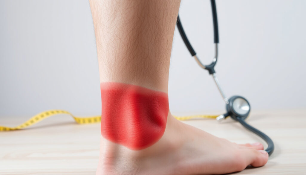
Unilateral swelling, redness or bluish‑purple discoloration, warmth, and visible veins. The leg may feel tight or heavy.
Early Signs Checklist
Swelling in one calf or ankle, pain or tenderness (often behind the knee), warmth, skin colour change, and a heavy or aching sensation.
Clot vs. Bruise Comparison
A bruise has a clear injury history, localised colour that fades, and normal skin temperature. DVT causes diffuse swelling, persistent warmth, and deep ache.
When to See a Doctor
Any one-sided leg swelling, pain, or colour change that does not improve with rest or elevation warrants prompt medical evaluation.
- Most visible DVT signs are non‑specific – redness, swelling, warmth – and can mimic other conditions.
- Pictures alone cannot diagnose DVT; imaging (ultrasound) is required.
- Early‑stage clots may not be visible on the skin surface; deep clots often have no external sign.
- Bruises and blood clots differ in shape, colour progression, and tenderness.
- Warmth and unilateral swelling are stronger clues than pain location.
- Symptoms that worsen or fail to improve with leg elevation are more concerning for DVT.
| Feature | Blood Clot (DVT) | Bruise |
|---|---|---|
| Color | Reddish, warm, skin may be tight | Black/blue/purple, fades to green/yellow |
| Shape | Diffuse swelling along vein | Irregular, well‑defined border |
| Pain | Deep ache, worsened by flexing foot | Tender to touch, localised |
| Temperature | Warm to touch | Normal skin temperature |
| Progression | Gradual swelling and redness | Fades over days to weeks |
How Does a DVT Appear on the Skin?
DVT‑related leg images typically show one‑sided swelling of the calf, ankle, foot, or behind the knee. The area may appear red, blue, or purple, and the skin often looks puffy or tense rather than simply bruised. Prominent surface veins may become more visible. Liv Hospital’s visual guide notes that the leg can feel warm or hot to the touch compared to the unaffected side.
What Do Early Stage Blood Clot Pictures Show?
Early‑stage images often reveal only mild swelling and a faint reddish or bluish tint. The changes may be subtle, especially when the clot is deep. The WebMD DVT slideshow illustrates that early signs include a feeling of heaviness or tightness, sometimes without visible colour change. Symptoms may come and go during the first 48 hours.
Ultrasound Imaging of Deep Vein Thrombosis
Duplex ultrasound is the standard imaging test for DVT. It uses sound waves to show blood flow and detect blockage. During a compression ultrasound, the doctor presses on the vein with the probe. A normal vein collapses; a clotted vein remains open and non‑compressible. The CDC confirms that ultrasound can show a thrombus directly and evaluate flow. For calf DVT, duplex sonography is considered the mainstay of imaging, according to Radiology Source.
What Do Removed Blood Clots Look Like?
Surgically removed clots vary in size and shape, from small gelatinous fragments to long, tubular casts of the vein. These images are typically used in medical education and are not helpful for self‑diagnosis. The American Society of Hematology provides treatment guidelines that mention thrombectomy and catheter‑directed thrombolysis for severe cases, but pictures of removed clots are rarely needed for patient decision‑making.
Early Signs and Symptoms of a Blood Clot in the Leg (With Comparisons)
Common Early Symptoms: Swelling, Pain, Redness
The most frequently reported early symptoms are unilateral swelling, pain or tenderness (often starting in the calf), warmth, and redness or discoloration. A feeling of heaviness, tightness, or aching in the limb is also common. According to the Mayo Clinic, these signs can develop gradually or appear suddenly.
How to Check for a Blood Clot at Home (Homan’s Test Caution)
The Homan’s test – flexing the foot upward to see if it causes calf pain – is considered unreliable and potentially dangerous. Forceful movement can dislodge a clot, leading to pulmonary embolism. The Society for Vascular Surgery strongly advises against relying on home tests and recommends professional evaluation if DVT is suspected.
Instead of self‑testing, note whether the swelling and pain improve with leg elevation. DVT symptoms often persist or worsen, while a pulled muscle or bruise tends to feel better after rest.
When to Suspect DVT vs. Other Conditions
DVT can resemble a sprain, strain, or superficial thrombophlebitis. If you have a history of recent surgery, prolonged immobility (e.g., long‑haul travel), pregnancy, or cancer, your risk is higher. The CDC emphasises that unilateral leg swelling and warmth are the most specific combination of signs. If both are present, medical imaging is warranted.
Blood Clot in Leg vs. Bruise: How to Tell the Difference
Key Visual Differences (Color, Texture, Size)
A bruise usually follows an injury and produces localised skin discoloration that changes over time – from black‑blue to green to yellow. The area is flat or slightly raised but not diffusely swollen. In contrast, DVT causes swelling of an entire segment of the leg (calf, ankle, or thigh), and the skin may appear red, blue, or purple with a tense, shiny appearance. Prominent veins may become more noticeable.
Pain Characteristics: Clot vs. Bruise
Bruise pain is typically tender to touch and localised directly over the discoloured area. DVT pain is described as a deep ache, often felt inside the calf or behind the knee. It may worsen when the foot is flexed upward (a motion that stretches the calf muscles) but, as noted earlier, this test is not reliable. The warmth of the skin over a clot is a key differentiating feature; a bruise does not generate heat.
Can a Bruise Turn into a Blood Clot?
A simple bruise does not become a DVT. However, a deep bruise combined with damage to a vein can theoretically trigger clotting. The Liv Hospital article explains that bruising and DVT can coexist, but they are separate processes. If a bruise is accompanied by significant swelling, warmth, or tenderness extending beyond the bruised area, a medical evaluation is recommended.
Treatment Options for Blood Clots in the Leg (With Procedure Images)
Medications: Anticoagulants and Thrombolytics
Anticoagulants, often called blood thinners, are the main treatment. They prevent the clot from growing and reduce the risk of pulmonary embolism. According to the WebMD slideshow, common medications include heparin, warfarin, and direct oral anticoagulants. In severe cases, thrombolytic drugs are used to dissolve the clot quickly.
Surgical Removal and Catheter Procedures (with images)
For large or high‑risk clots, procedures such as catheter‑directed thrombolysis or mechanical thrombectomy may be performed. These interventions are shown in medical literature but are not typically used for first‑line treatment. The Society for Vascular Surgery notes that surgery is reserved for cases where the clot threatens the limb or when anticoagulation is contraindicated.
Lifestyle Changes and Compression Therapy
Compression stockings can reduce leg swelling and the heaviness associated with DVT. Elevating the leg above heart level also helps improve blood flow and decrease pain. The SVS guidelines recommend compression therapy after the acute phase to prevent post‑thrombotic syndrome.
Staying active, hydrating, and wearing compression stockings during long travel can also help prevent future clots.
When to Seek Emergency Care for a Leg Blood Clot
Red Flags: Sudden Severe Pain, Shortness of Breath, Chest Pain
If a DVT breaks off and travels to the lungs, it can cause a pulmonary embolism – a life‑threatening emergency. Sudden shortness of breath, chest pain that worsens with deep breathing, coughing up blood, or a rapid heart rate require immediate medical attention. The Mayo Clinic stresses that prompt treatment of a leg clot drastically reduces the risk of embolism.
How Quickly Does a DVT Progress?
The timeline varies widely. Some clots cause noticeable symptoms within hours; others remain silent for days or weeks. The CDC notes that untreated DVT can lead to pulmonary embolism at any stage. Early detection is key.
Importance of Prompt Diagnosis and Imaging
Doppler ultrasound can be performed quickly in an emergency department. The procedure is non‑invasive and provides immediate results. Delaying evaluation increases the risk of clot extension and complications.
How Does a Blood Clot in the Leg Progress Over Time?
The visual and symptomatic progression of untreated DVT follows a general pattern, though many clots remain asymptomatic. The following timeline is based on clinical observations from the Society for Vascular Surgery and other sources.
- Day 1–2: Mild pain or tenderness, slight swelling. Skin may appear normal or faintly pink. Some people experience only a feeling of heaviness.
- Day 3–7: Swelling becomes more noticeable. Skin turns red or bluish and feels warm. Pain worsens, especially when walking or flexing the foot.
- Week 2+: Without treatment, the leg may look visibly larger. Skin may become shiny, tight, and discoloured (brownish or purplish). Risk of pulmonary embolism is highest during this period.
- Beyond weeks: Chronic changes can develop, including post‑thrombotic syndrome with persistent swelling, skin ulcers, and pain.
Important: Many DVTs cause no visible symptoms at any stage. The absence of leg changes does not rule out a clot.
Can You Rely on Pictures Alone to Diagnose a Blood Clot?
Visual identification has limited reliability. Even experienced clinicians use ultrasound to confirm DVT because symptoms and appearance overlap with many other conditions.
Established Information
- Unilateral swelling, warmth, and redness are the most typical visible signs.
- Ultrasound (duplex or compression) is the gold‑standard diagnostic tool.
- D‑dimer blood testing can help decide if imaging is needed but cannot confirm DVT alone.
Information That Remains Unclear
- Skin colour changes can be caused by infection, injury, or superficial thrombosis.
- Half of all DVTs cause no visible symptoms.
- No single photograph can reliably distinguish DVT from a muscle strain or cellulitis.
The Liv Hospital article and the CDC both advise against self‑diagnosis based on pictures. If you suspect a clot, seek medical evaluation.
Why Is There So Much Concern About Blood Clots in the Leg?
Deep vein thrombosis affects an estimated 1–2 per 1,000 adults each year. Risk factors include surgery, prolonged immobility (e.g., long flights), pregnancy, cancer, and inherited clotting disorders. The condition is serious because untreated DVT can lead to pulmonary embolism, a leading cause of preventable hospital death.
Public anxiety is understandable – many people want to spot the problem early. Yet the desire for self‑assessment can lead to over‑reliance on visual clues. The American Society of Hematology emphasises that education about symptoms must be paired with a clear message: only imaging can provide a definitive answer. For those looking to understand more about complex vascular conditions, the Twin to Twin Transfusion Syndrome Guide covers another serious condition involving blood flow.
Understanding the limitations of visual identification helps reduce unnecessary worry while ensuring that true emergencies are not missed.
Where Can You Find Reliable Information and Images?
Several authoritative organizations provide free, evidence‑based visuals and guidance. The following are among the most trusted.
“Deep vein thrombosis can occur without any symptoms. If you have swelling, pain, and warmth in your leg that doesn’t go away, see your doctor.”
– Centers for Disease Control and Prevention
“Ultrasound is the most common and reliable way to diagnose DVT. It uses sound waves to create images of blood flow.”
“Blood thinners are the main treatment for DVT. They prevent the clot from getting bigger and reduce the risk of a pulmonary embolism.”
– American Society of Hematology
For those seeking additional reading, the Luton and Dunstable Hospital Guide provides general healthcare navigation advice, while the Twin to Twin Transfusion Syndrome Guide covers another serious vascular condition.
Key Takeaways About Blood Clot in Leg Pictures
Pictures can help you recognise the possible signs of a DVT – especially one‑sided swelling, warmth, and colour change – but they cannot replace a medical diagnosis. If you suspect a blood clot, do not massage the leg; keep it elevated and seek prompt evaluation. Early treatment with anticoagulants is highly effective. Prevention through movement, hydration, and compression during long travel remains the best strategy.
Frequently Asked Questions
Can a blood clot in the leg disappear on its own?
Small clots may be reabsorbed by the body, but untreated DVTs can break off and cause pulmonary embolism. Medical treatment is recommended.
Are blood clots in the leg always painful?
No, many DVTs are painless, especially if deep. Pain often occurs when the clot is large or close to the skin.
How accurate is home examination for DVT?
Home tests (e.g., Homan’s sign) are unreliable and can dislodge a clot. Always seek professional evaluation.
What does a blood clot look like on ultrasound?
Ultrasound shows a hypoechoic area within the vein, lack of compressibility, and absent blood flow. Images are available in the ultrasound section above.
Can exercise dislodge a blood clot?
Yes, vigorous exercise or massage can dislodge a clot. Rest and immobilisation are advised until treatment begins.
Is it safe to take a flight after a DVT diagnosis?
Air travel shortly after diagnosis is generally not recommended. Your doctor will advise when it is safe, often after a few weeks of treatment.
What is the difference between a blood clot and superficial thrombophlebitis?
Superficial thrombophlebitis affects veins near the skin surface and usually causes a hard, tender cord. DVT involves deeper veins and carries a higher risk of embolism.
Do compression stockings cure DVT?
Compression stockings do not dissolve the clot, but they reduce swelling and help prevent post‑thrombotic syndrome. They are used alongside anticoagulants.
How long does it take to recover from a DVT?
Recovery varies. Most people take anticoagulants for 3–6 months. Swelling and discomfort may take weeks to resolve, and some have lasting symptoms.
More related posts
 Swimming Pools Near Me – Public Pools, Hours, Costs Guide
 Wake Up Dead Man – Cast, Killer and Netflix Release Guide
 Barbara Knox – Age, Net Worth, and Life in 2025
 Liverpool Water Street Incident – 130 Injured in Parade Attack
 Fabrizio Romano Man Utd – Tchouameni, Ederson and Ugarte Updates
 Chris Briney – Age, Height, Parents and Girlfriend Revealed
 7 Wonders of the World – Complete List, History and Facts
 Where Is Virgin River Filmed – Complete BC Locations Guide
